amplifyeyecare-logo-base2-icon

Scleral Lenses & Other Treatments for Corneal Ectasia

Scleral lenses offer a breakthrough in corneal ectasia management. Learn how you can benefit from this innovative treatment.

Find an amplify doctor near you
Scleral Lenses & Other Treatments for Corneal Ectasia Optometrist
Table of Contents

According to a study published in the Journal of Cataract & Refractive Surgery, the incidence rate of corneal ectasia following LASIK surgery, one of its common causes, is estimated to be around 0.2 to 0.6 percent in the United States. This article is covering corneal ectasia that is a result of a complication of certain medical procedures, such as refractive surgery or corneal cross-linking. There is another kind of ectasia, which is not the subject of this article, in patients with keratoconus in which the cornea thins and weakens, leading to bulging and distortion.

The average age of onset for post-LASIK ectasia is generally within the first 1-2 years after surgery. Diagnosis of corneal ectasia is often delayed due to its similarity to other eye conditions like myopia and astigmatism. Early detection and treatment of corneal ectasia can prevent severe visual impairment.

Corneal ectasia is a condition that particularly stands out due to its complex nature. It is a progressive eye disorder that causes the cornea, the clear front surface of the eye, to thin and bulge outwards, leading to a distortion of vision.

Amplify EyeCare

The Gold Standard in the Treatment of Corneal Ectasia

For many years, the treatment of corneal ectasia has been a challenge for many eye care professionals. However, thanks to modern eye care advancements, the landscape of corneal ectasia treatment has drastically changed for the better.

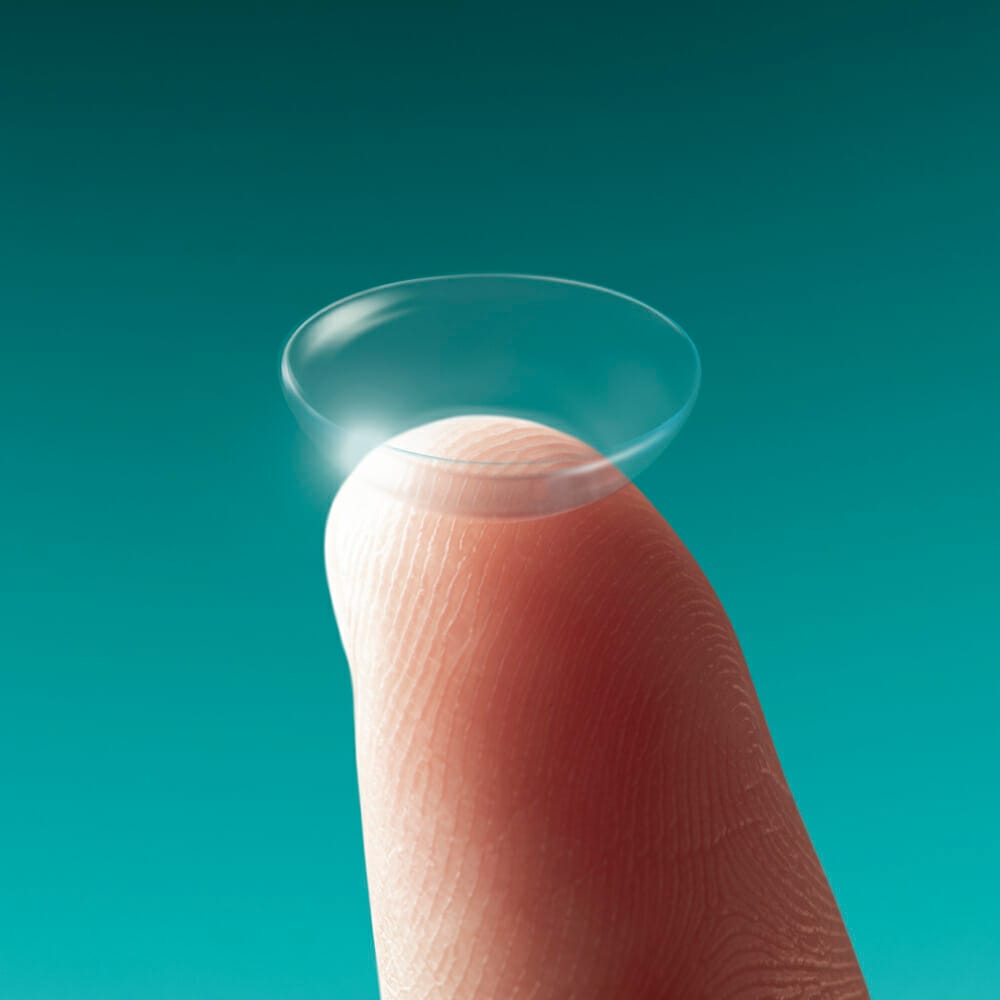
We specialize in fitting advanced, medically necessary contact lenses, specifically custom scleral lenses. These lenses have emerged as the gold standard in the treatment of corneal ectasia and other corneal irregularities. Unlike traditional contact lenses, scleral lenses are designed to vault over the damaged ocular surface and create a new undamaged surface. They not only correct vision but also promote ocular surface healing and comfort.

Benefits of Scleral Lenses

One of the significant benefits of scleral lenses is that they create a pocket filled with saline, which keeps the eyes moist and comfortable all day long. These lenses can:

  • Improve the clarity of your vision
  • Reduce the need for corneal transplantation
  • Protect the cornea from the environment and the lids
  • By utilizing scleral lenses, you can mitigate the effects of corneal ectasia and experience an improved quality of life.

Fitting Scleral Lenses for Corneal Ectasia

Our fitting process involves several crucial steps to ensure maximum comfort, optimal fit, and excellent vision correction.

Comprehensive Eye Examination: We begin with a thorough eye examination to understand the precise shape and health of your cornea, along with assessing your overall ocular health.

Specialized Imaging: We employ state-of-the-art corneal imaging techniques like corneal topography and optical coherence tomography (OCT) to map the surface of your eyes. These images allow us to design a lens that perfectly fits your unique eye shape.

Custom Lens Design: Based on the detailed measurements and your specific needs, we design a custom scleral lens. These lenses vault over the cornea, resting on the sclera (the white part of the eye), and create a new, smooth optical surface.

Trial Lens Fitting and Assessment: Next, we fit you with a trial lens to assess the comfort and fit. We may make necessary adjustments to the lens design based on this trial fit.

Instruction and Follow-up: Once the final lens design is confirmed and your lenses are ready, we will teach you how to correctly insert, remove, and care for your scleral lenses. Follow-up appointments are scheduled to ensure your lenses are performing well and that you are comfortable wearing them.

What Causes Corneal Ectasia

What Causes Corneal Ectasia

Corneal ectasia often develops as a complication of LASIK or PRK eye surgery, but it can also be associated with a genetic disorder known as keratoconus. As such, the terms "keratoconus and corneal ectasia" are often used interchangeably in certain contexts, although they have distinct causes. Predisposing factors like high myopia, young age, and low residual stromal bed thickness increase the risk of post-LASIK ectasia. Environmental factors, such as eye rubbing, have been linked to the progression of corneal ectasia.

What Causes Corneal Ectasia

What Causes Corneal Ectasia

Corneal ectasia often presents with a range of symptoms which can progress gradually over time. The presentation can vary from person to person and the severity of symptoms can also differ.

However, starting from the most common symptoms:

  • Blurred Vision: This is often the first symptom that patients notice. It's due to the irregular shape of the cornea causing light to be improperly focused on the retina.
  • Progressive Near-sightedness (Myopia): Patients often find that they are becoming more near-sighted, which is often uncorrectable with glasses.
  • Astigmatism: This is a common symptom in which patients experience distorted vision, due to the irregularly shaped cornea.
  • Increased Light Sensitivity: Many patients experience increased sensitivity to light and glare, which can become more severe in bright conditions.
  • Halos and Ghosting: Some patients may start to see halos around lights, or ghosting (double images), especially at night.
  • Sudden worsening or clouding of vision: This can occur due to acute corneal hydrops, a complication where fluid accumulates within the cornea.
  • Eye discomfort or pain: While not as common as visual disturbances, some patients may experience discomfort or a foreign body sensation in the eye.
  • Headaches: These can occur due to the strain of trying to focus with distorted vision.
  • Eye redness: This can be a response to the discomfort or the foreign body sensation in the eye.
  • Difficulty with night vision: The irregular corneal surface can make seeing at night or in dim light difficult.
  • Increased difficulty reading: Due to the changes in vision, reading might become more difficult over time.
  • Frequent changes in eyeglass prescription: As the shape of the cornea changes, so does the need for different corrective lenses.
  • Difficulty driving: This can be due to a combination of visual symptoms such as glare, halos, and difficulty with night vision.
  • Trouble recognizing faces: In more severe cases, visual distortion can reach the point where recognizing faces is difficult.
  • Inability to wear contact lenses: As the condition progresses, the cornea can become too irregular for standard contact lenses to fit comfortably or effectively.
  • Tearing or watery eyes: Some people may have increased tearing due to eye irritation.
  • Intolerance to bright light (Photophobia): Some patients experience significant discomfort or difficulty seeing in bright lights.
  • Decreased visual acuity: A gradual decline in how much detail can be discerned visually.
  • Eye strain: Trying to focus vision may lead to noticeable eye strain over time.
  • Swelling of the eye or eyelid: This is more rare, but can occur, especially during episodes of acute corneal hydrops.
Find an amplify doctor near you
Other Treatment Options for Corneal Ectasia

Other Treatment Options for Corneal Ectasia

Corneal ectasia requires comprehensive management to mitigate its symptoms and halt its progression. While scleral lenses are indeed a prominent treatment modality, various other therapeutic strategies exist to cater to different stages and severity levels of corneal ectasia. Let's explore some of these alternatives.

Corneal Collagen Cross-linking (CXL)

A very effective treatment, corneal collagen cross-linking (CXL), strengthens the cornea by creating new links between its collagen fibers. This treatment can halt the progression of corneal ectasia and, in some cases, may even lead to a partial reversal of the condition. In many cases cross linking will be done in combination with scleral lenses for the best outcome. 

Rigid Gas Permeable Contact Lenses

Another popular choice for patients with corneal ectasia is rigid gas permeable (RGP) contact lenses. They provide a smooth front surface to the eye, correcting vision problems caused by an irregularly shaped cornea. However, they may be less comfortable than scleral lenses, especially for sensitive eyes.

Hybrid Lenses

Hybrid lenses combine a rigid gas permeable center with a soft lens skirt, providing the crisp vision of RGPs and the comfort of soft lenses. They can be a good choice for patients who struggle with wearing traditional RGPs.

Intacs

Intacs are tiny plastic inserts that are surgically placed in the cornea to reshape it for clearer vision. They can be a good option for patients who cannot achieve suitable vision with contact lenses or glasses but are not candidates for corneal transplant.

Corneal Transplant

In severe cases, a corneal transplant may be required. This involves removing the central part of the cornea and replacing it with donor tissue. While effective, a corneal transplant is generally considered as a last resort due to the associated risks and lengthy recovery time.

The National Eye Institute reports that the FDA approval of Corneal Collagen Cross-Linking in 2016 has provided a promising treatment option for halting the progression of conditions leading to corneal ectasia, thereby potentially reducing the number of corneal transplants needed.

Keratoconus vs Corneal Ectasia

Keratoconus and corneal ectasia share similarities but are distinct conditions. Understanding the differences between the two can help clarify their unique characteristics and treatment options.

Keratoconus is a naturally occurring degenerative condition. It typically begins in the teenage years or early twenties, causing the cornea to thin and bulge into a cone-like shape. This distortion can lead to blurred and distorted vision. Keratoconus can progress for 10-20 years before it slows down and stabilizes.

Corneal ectasia, on the other hand, is usually an iatrogenic condition, meaning it's caused by medical intervention, specifically refractive surgery like LASIK or PRK. Post-LASIK ectasia follows a pattern similar to keratoconus but usually occurs months to years after surgery.

Although both conditions share common symptoms such as blurred or distorted vision, increased sensitivity to light, and difficulty with night vision, they differ in their causes and progression rates. Their treatment methods are also similar, often involving custom contact lenses or corneal cross-linking to strengthen the cornea and improve vision. However, it's crucial to consult with our optometrist to determine the best course of action for each unique case.

What Causes Corneal Ectasia
Specialty Contacts optometry and eye care
Amplify EyeCare and Optometrists

Find an Amplify Practice in Your Area

Amplify EyeCare is a team of a passionate and experienced optometrists practicing eye care at the cutting edge of technology and vision science. We are growing with new locations coming across the US.
Visit a Specialty Contacts eye doctor at an Amplify EyeCare practice near you:

Contact Us To Amplify Your EyeCare

arrow-uparrow-right